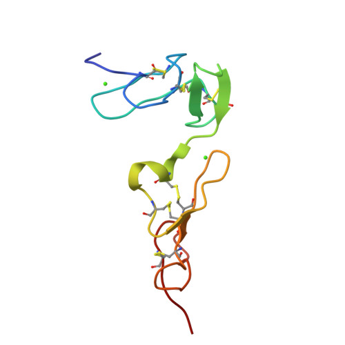
Vitamin K-dependent protein S
Homo sapiensTaxon: 9606
676
amino acids
75.1 kDa
theoretical
1
PDB entries
2
recorded
Function
Localization & Distribution
Tissue Specificity
Plasma
Related Diseases
Thrombophilia due to protein S deficiency, autosomal dominant
A hemostatic disorder characterized by impaired regulation of blood coagulation and a tendency to recurrent venous thrombosis. Based on the plasma levels of total and free PROS1 as well as the serine protease-activated protein C cofactor activity, three types of THPH5 have been described: type I, characterized by reduced total and free PROS1 levels together with reduced anticoagulant activity; type III, in which only free PROS1 antigen and PROS1 activity levels are reduced; and the rare type II which is characterized by normal concentrations of both total and free PROS1 antigen, but low cofactor activity.
Thrombophilia due to protein S deficiency, autosomal recessive
A very rare and severe hematologic disorder resulting in thrombosis and secondary hemorrhage usually beginning in early infancy. Some affected individuals develop neonatal purpura fulminans, multifocal thrombosis, or intracranial hemorrhage.
Amino Acid Sequence
MRVLGGRCGALLACLLLVLPVSEANFLSKQQASQVLVRKRRANSLLEETKQGNLERECIE
ELCNKEEAREVFENDPETDYFYPKYLVCLRSFQTGLFTAARQSTNAYPDLRSCVNAIPDQ
CSPLPCNEDGYMSCKDGKASFTCTCKPGWQGEKCEFDINECKDPSNINGGCSQICDNTPG
SYHCSCKNGFVMLSNKKDCKDVDECSLKPSICGTAVCKNIPGDFECECPEGYRYNLKSKS
FASTA format · 676 amino acids · mass 75.1 kDa
Experimental Structures
1 PDB entriesPost-Translational Modifications
- •The iron and 2-oxoglutarate dependent 3-hydroxylation of aspartate and asparagine is (R) stereospecific within EGF domains