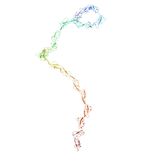
Complement factor H
Homo sapiensTaxon: 9606
1231
amino acids
139.1 kDa
theoretical
48
PDB entries
4
recorded
Function
Localization & Distribution
Tissue Specificity
Expressed in the retinal pigment epithelium (at protein level) (PubMed:25136834). CFH is one of the most abundant complement components in blood where the liver is the major source of CFH protein in vivo. in addition, CFH is secreted by additional cell types including monocytes, fibroblasts, or endothelial cells (PubMed:2139673, PubMed:25136834, PubMed:2968404, PubMed:6444659)
Related Diseases
Basal laminar drusen
Drusen are extracellular deposits that accumulate below the retinal pigment epithelium on Bruch membrane. Basal laminar drusen refers to an early adult-onset drusen phenotype that shows a pattern of uniform small, slightly raised yellow subretinal nodules randomly scattered in the macula. In later stages, these drusen often become more numerous, with clustered groups of drusen scattered throughout the retina. In time these small basal laminar drusen may expand and ultimately lead to a serous pigment epithelial detachment of the macula that may result in vision loss.
Complement factor H deficiency
A disorder that can manifest as several different phenotypes, including asymptomatic, recurrent bacterial infections, and renal failure. Laboratory features usually include decreased serum levels of factor H, complement component C3, and a decrease in other terminal complement components, indicating activation of the alternative complement pathway. It is associated with a number of renal diseases with variable clinical presentation and progression, including membranoproliferative glomerulonephritis and atypical hemolytic uremic syndrome.
Hemolytic uremic syndrome, atypical, 1
An atypical form of hemolytic uremic syndrome. It is a complex genetic disease characterized by microangiopathic hemolytic anemia, thrombocytopenia, renal failure and absence of episodes of enterocolitis and diarrhea. In contrast to typical hemolytic uremic syndrome, atypical forms have a poorer prognosis, with higher death rates and frequent progression to end-stage renal disease.
Macular degeneration, age-related, 4
A form of age-related macular degeneration, a multifactorial eye disease and the most common cause of irreversible vision loss in the developed world. In most patients, the disease is manifest as ophthalmoscopically visible yellowish accumulations of protein and lipid that lie beneath the retinal pigment epithelium and within an elastin-containing structure known as Bruch membrane.
Amino Acid Sequence
MRLLAKIICLMLWAICVAEDCNELPPRRNTEILTGSWSDQTYPEGTQAIYKCRPGYRSLG
NVIMVCRKGEWVALNPLRKCQKRPCGHPGDTPFGTFTLTGGNVFEYGVKAVYTCNEGYQL
LGEINYRECDTDGWTNDIPICEVVKCLPVTAPENGKIVSSAMEPDREYHFGQAVRFVCNS
GYKIEGDEEMHCSDDGFWSKEKPKCVEISCKSPDVINGSPISQKIIYKENERFQYKCNMG
FASTA format · 1231 amino acids · mass 139.1 kDa
Experimental Structures
48 PDB entriesPost-Translational Modifications
- •Sulfated on tyrosine residues
- •According to a report, Asn-217 is not glycosylated (PubMed:17591618). Another study observed glycosylation at this position (PubMed:19139490)