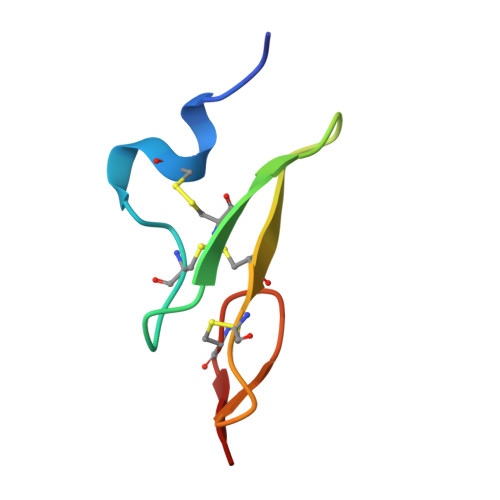
P08709
FA7_HUMANF7
Coagulation factor VII
Homo sapiensTaxon: 9606
3D-structureAlternative splicingBlood coagulationCalciumCleavage on pair of basic residuesDirect protein sequencingDisease variantDisulfide bondEGF-like domainGamma-carboxyglutamic acidGlycoproteinHemostasis+11
Sequence Length
466
amino acids
Molecular Weight
51.6 kDa
theoretical
Experimental Structures
50
PDB entries
Related Diseases
1
recorded
Function
Initiates the extrinsic pathway of blood coagulation. Serine protease that circulates in the blood in a zymogen form. Factor VII is converted to factor VIIa by factor Xa, factor XIIa, factor IXa, or thrombin by minor proteolysis. In the presence of tissue factor and calcium ions, factor VIIa then converts factor X to factor Xa by limited proteolysis. Factor VIIa also converts factor IX to factor IXa in the presence of tissue factor and calcium (PubMed:271951)
Localization & Distribution
Tissue Specificity
Plasma
Related Diseases
Factor VII deficiency
A hemorrhagic disease with variable presentation. The clinical picture can be very severe, with the early occurrence of intracerebral hemorrhages or repeated hemarthroses, or, in contrast, moderate with cutaneous-mucosal hemorrhages (epistaxis, menorrhagia) or hemorrhages provoked by a surgical intervention. Finally, numerous subjects are completely asymptomatic despite very low factor VII levels.
Amino Acid Sequence
MVSQALRLLCLLLGLQGCLAAGGVAKASGGETRDMPWKPGPHRVFVTQEEAHGVLHRRRR
ANAFLEELRPGSLERECKEEQCSFEEAREIFKDAERTKLFWISYSDGDQCASSPCQNGGS
CKDQLQSYICFCLPAFEGRNCETHKDDQLICVNENGGCEQYCSDHTGTKRSCRCHEGYSL
LADGVSCTPTVEYPCGKIPILEKRNASKPQGRIVGGKVCPKGECPWQVLLLVNGAQLCGG
FASTA format · 466 amino acids · mass 51.6 kDa
Experimental Structures
50 PDB entries1BF91CVW1DAN1DVA1F7E1F7M1FAK1FF71FFM1J9C1JBU1KLI1KLJ1O5D1QFK1W0Y1W2K1W7X1W8B1WQV1WSS1WTG1WUN1WV71YGC1Z6J2A2Q2AEI2AER2B7D2B8O2BZ62C4F2EC92F9B2FIR2FLB2FLR2PUQ2ZP02ZWL2ZZU3ELA3TH23TH33TH44IBL4ISH4ISI4JYU
Post-Translational Modifications
- •The vitamin K-dependent, enzymatic carboxylation of some glutamate residues allows the modified protein to bind calcium
- •The iron and 2-oxoglutarate dependent 3-hydroxylation of aspartate and asparagine is (R) stereospecific within EGF domains
- •O- and N-glycosylated. N-glycosylation at Asn-205 occurs cotranslationally and is mediated by STT3A-containing complexes, while glycosylation at Asn-382 is post-translational and is mediated STT3B-containing complexes before folding. O-fucosylated by POFUT1 on a conserved serine or threonine residue found in the consensus sequence C2-X(4,5)-[S/T]-C3 of EGF domains, where C2 and C3 are the second and third conserved cysteines
- •Can be either O-glucosylated or O-xylosylated at Ser-112 by POGLUT1 in vitro